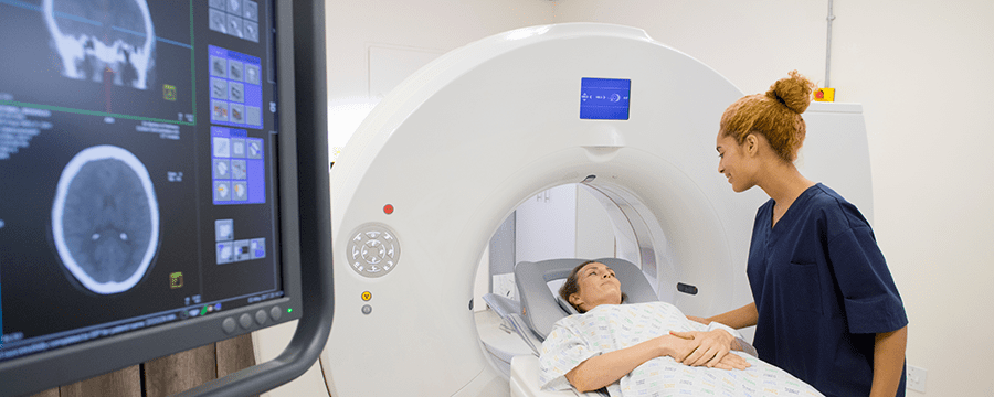
From mammograms to CT scans, MRIs and much more, HaysMed offers a full array of imaging services in Hays and through our mobile imaging clinics across the region.

Advanced Imaging Services for Expert Diagnostics and Care
At HaysMed Imaging Center, we provide expert diagnostic imaging services, both inpatient and outpatient. We also extend our ultrasound services to the northwestern part of Kansas via our Mobile Imaging Services. Our board-certified, state-licensed staff combines highly advanced imaging technology, along with their specialized training to meet all your imaging needs.
Imaging Services
At our Imaging Center, we provide:
- Diagnostic imaging (X-ray) including interventional procedures
- Computerized Axial Tomography (CT) using two multi detector scanners with ASIR to reduce radiation exposure
- Nuclear medicine, including diagnostic, cardiac and PET/CT imaging
- Magnetic resonance imaging (MRI): ACR-certified in MRA, MSK, body, brain, and spine
- MRI breast: ACR-certified and breast biopsy
- MRI prostate imaging for fusion capability with urology
- MRI treatment planning for radiation oncology.
- MRI enterography
- Ultrasound including breast, breast biopsy, abdominal, vascular and OB
- Ultrasound echocardiography (mobile only)
- ACR-certified breast stereo biopsy
- ACR-certified digital mammography screening/diagnostic with tomography
- DXA (bone mineral analysis)
Diagnostic X-rays and MRIs are also available at the HaysMed Orthopedic Institute. Visit our Imaging Services page for more details about select imaging procedures.
HaysMed Imaging Center is located on the main hospital campus, with a satellite location in the HaysMed Orthopedic Institute.
HaysMed Imaging Center
2220 Canterbury Drive
Hays, Kansas
785-623-5701
HaysMed Orthopedic Institute
2500 Canterbury, Suite 112
Hays, Kansas
785-261-7560
Please note: HaysMed is a teaching facility. A teaching hospital partners with medical and nursing schools and education programs to improve health care through learning. As part of the training and education process, medical students may be part of your health care team. These students provide patient care under the direct supervision of the radiologist, radiologic technologist and clinical supervisors, who are all registered, licensed and responsible for training the next generation of health care providers.
Advanced Imaging Services
When you are relying on your doctor to make an accurate diagnosis, image quality is everything. HaysMed Imaging Center offers the latest technological advances along with standard imaging modalities used for diagnostic, interventional and therapeutic purposes.
X-ray studies
X-rays use low doses of radiation and are often performed as the initial examination to diagnose a wide variety of diseases and injuries. Radiography with X-ray is the starting point for diagnosing or screening of a variety of health issues, including cancer, pneumonia, emphysema, and fractures. The new digital imaging equipment provides better and clearer images with high resolution and reduced radiation. It is easily available to the medical community with 24/7 availability, quickly giving radiologists and providers the ability to better see disease processes at their earliest stages.
Mammography
For advanced breast images to catch and detect breast cancer and other breast abnormalities, HaysMed uses 3D mammography. Learn more here.
Fluoroscopy
Fluoroscopy is another type of X-ray technology which is used to produce real-time images of internal body anatomy on a video screen, even while the movements are occurring. Images of diaphragm motion, swallowing coordination, and a barium-filled gastrointestinal tract can be recorded to be reviewed and evaluated at a later time.
Spiral Computed Tomography
Computed tomography is a highly advanced X-ray procedure that combines X-rays with computers to produce two-dimensional cross-section images of organs and other body parts. A CT scan is useful in evaluating the brain, spine, chest, abdomen, and pelvis to determine the site, type and extent of disease or injury. CT can determine if internal bleeding is occurring or visualize the size and extent of tumors. Spiral CT is the latest technology, particularly used during evaluation of cancer and trauma patients.
Magnetic Resonance Imaging (MRI)
Magnetic Resonance Imaging produces three-dimensional visual images of internal structures in any plane using strong electromagnetic fields and powerful computers. In MRI, the patient is placed inside a powerful magnet that aligns the water molecules contained in the body tissues. A radio signal is directed to the body part being examined, temporarily disrupting this alignment. When the radio signal stops, the water molecules return to alignment at different rates and emit signals that are received and processed at a computer, which converts the data to a series of images. MRI is currently the most sensitive, non-invasive way of imaging the brain, spine, soft tissues, organs, and musculoskeletal structures, as well as breast and prostate.
MR Angiography is a special technique used to process MRI information of the blood vessels to diagnose abnormalities, aneurysm, occlusions, and ruptures.
Sono/Ultrasound
Sono/Ultrasound produces images using high-frequency sound waves rather than X-rays. Sound waves are directed into the body from a transducer through the skin surface. When these sound waves come into contact with the internal structures, part of the sound energy is reflected back to the body surface, to the transducer where it is converted electronically into an image.
Since there is no X-ray involved, Ultrasound is commonly used during pregnancy to determine fetal position, age, and gross anatomy. It is also used to evaluate numerous other structures such as the gallbladder, spleen, liver, kidneys, and thyroid.
Doppler Ultrasound detects blood flow in the veins and arteries and is used to evaluate and diagnose problems such as blood clots, venous reflux disease, pre-operative mapping of vessels, and atherosclerotic disease.
Nuclear Medicine
Nuclear medicine involves using radioactive isotopes to image different organ systems in the body, including brain, thyroid, lungs, heart, liver, kidneys, gallbladder, bones, and tumor imaging for cancer diagnosis. Nuclear medicine differs from other imaging because it uses gamma rays instead of X-rays and it assesses the function of the body instead of the anatomy. Cardiac stress testing is a common nuclear medicine test to look at the perfusion of blood to the heart and rule out ischemia.
PET/CT is a form of nuclear medicine, and a mobile service offers PET/CT one day each week.
Interventional Radiology/Special Procedures
Interventional Radiology/Special Procedures refers to a number of different procedures involving catheters, contrast media (dye), stents, drains, and needles placed through the skin surface into the body, without surgical intervention. These diagnostic and therapeutic techniques are often outpatient procedures and are generally less invasive than the corresponding surgical procedures:
- Needle biopsy. Thin needles, or core biopsy devices, can be introduced through the skin to sample tissue from within the body, without the need for a surgical incision. CT, fluoroscopic or ultrasound guidance permits the samples of the appropriate organ or mass to be obtained for subsequent evaluation by a pathologist. Some breast specimens are retrieved using a digital stereotactic biopsy device.
- Stereotactic biopsy. This type of biopsy is a major advance in the diagnosis of breast cancer, which is now available at HaysMed. Developed over the last 10 years, stereotactic biopsy is a procedure of computerized directed needle biopsy of the breast which provides tissue diagnosis of mammographic findings without surgery. Pathology results are available in two days.
- Abscess Drainage. Abscesses can, in some cases, be drained without surgical incision, again using CT, fluoroscopic, or ultrasound guidance. A drain tube is left in place, open to the skin surface, while the abscess resolves.
HaysMed Imaging Center is located on the main hospital campus, with a satellite location in the HaysMed Orthopedic Institute.
HaysMed Imaging Center
2220 Canterbury Drive
Hays, Kansas
785-623-5701
HaysMed Orthopedic Institute
2500 Canterbury, Suite 112
Hays, Kansas
785-261-7560
For information contact us today.
Mammography (Breast Imaging)
Located at The Breast Care Center
Park in parking lot C and use entrance C
Screening and diagnostic mammography, along with self-examination and a clinical breast exam, are powerful weapons for early detection of breast cancer. Breast cancer is most treatable when caught early.
Our 3D Mammography technology is an advanced system that captures three-dimensional images of the internal structures of the breast. It allows the radiologist to scroll through the breast to find cancer or other breast abnormalities at an earlier stage than 2D mammography. Our mammography services at HaysMed are accredited by the American College of Radiology and certified by the U.S. Food and Drug Administration.
Breast tomosynthesis is the latest technology in the fight against breast cancer, enabling the three dimensional imaging needed to identify breast cancer sooner, when it’s smaller and less likely to have spread to other parts of the body. Breast tomosynthesis is used in conjunction with digital mammography as part of your annual screening mammogram to capture more breast images. Using breast tomosynthesis and digital mammography together for screening has been proven to reduce “call-backs.”
Breast tomosynthesis converts digital breast images into a stack of very thin layers or “slices” – building what is essentially a three-dimensional mammogram. This type of breast imaging allows doctors to examine breast tissue one layer (or millimeter) at a time, instead of viewing all the complexities of your breast tissue in a flat image. Fine details are more clearly visible, no longer hidden by the tissue above and below.
A tomosynthesis exam is very similar to a traditional mammogram. The exam time is approximately the same as a digital mammogram and delivers a similar radiation dose.
Just as with your digital mammogram, your breast will be compressed while the X-ray arm makes a quick arc over the breast taking a series of images at a number of angles. Both 2D and 3D images are obtained simultaneously. With tomosynthesis, additional compression and adjustment of the breast is not required.
Its advantages include:
- 30% increase in breast cancer detection
- Less time needed for breast compression during testing
- Lower average radiation dose
- Quality images viewed quickly to assure the evaluation is complete prior to the patient leaving the room
- May provide a lower chance you will have to come back for additional mammographic imaging
Should you consider 3D mammography?
Ask yourself the following questions. If you answer yes to any of these, 3D mammography may be right for you. Talk to your doctor about it today.
- Do you have dense breasts?
- Do you have a history of call backs with your mammograms?
- Do you have a family history of call backs with your mammograms?
- Do you have a history of breast abnormalities or cancer?
Mammograms are conducted at:
HaysMed Breast Care Center
2220 Canterbury Drive
Hays, Kansas
You can schedule a mammogram by:
-You can call 785-623-6565
-If you have a patient portal account, you can log in and schedule an appointment. https://rb.gy/cfjfhj
-If you don’t have a patient portal account you can create one and then schedule your mammogram. https://rb.gy/cfjfhj
If you have questions about mammograms, please call the Breast Care Center at 785-623-5114 or talk with your healthcare provider.
HaysMed Mobile Imaging Network
HaysMed has a long history of sharing technology and technical expertise with healthcare practitioners in western Kansas. We bring imaging technologies that rural hospitals alone could not afford to these critical access hospitals to provide the essential diagnostic imaging services and keep patients connected to their local, family doctor, close to home.
Mobile imaging services from HaysMed include ultrasound with color doppler. Nuclear Medicine is limited to a stationary service at Pawnee Valley Community Hospital. All mobile imaging services are supervised by the experts of Radiology Associates of HaysMed and/or the cardiologists at DeBakey Heart Institute (for cardiac imaging).
Our mobile technical staff members are credentialed in their specialties and participate in continuing education, so we continue to offer the latest techniques for best practice.
For nuclear medicine, including cardiac stress testing, contact centralized scheduling at 785-623-6565. Appointments are available weekly in Larned.
For mobile echo only, contact your local radiology department for an appointment. Appointments are available weekly in Phillipsburg at the Phillips County Health System.
For mobile ultrasound, echo, vascular, contact your local radiology department for an appointment. Appointments are available in the following locations:
Atwood at the Rawlins County Health Center
Dighton at Lane County Hospital
Hill City at Graham County Hospital
Larned at Pawnee Valley Community Hospital
Osborne at Osborne County Memorial Hospital
Ransom at Grisell Memorial Hospital District #1 –
Bone Density Testing
Located at The Breast Care Center
Park in parking lot C and use entrance C
DEXA (dual energy X-ray absorptiometry) is used to determine your risk for bone fractures. It uses a small amount of radiation to look at the density of your bones and mineral content. Scans are safe, easy and determine if you are at risk for osteoporosis. Knowing if you have osteoporosis is important as there are treatment options available to minimize the effects of this process.
For more information about bone density testing, talk to your primary care provider.
Our Locations
The HaysMed Radiology is located on the main hospital campus, with a satellite location in the HaysMed Orthopedic Institute.
HaysMed Radiology
2220 Canterbury Drive
Hays, KS
785-623-5701
HaysMed Orthopedic Institute
2500 Canterbury, Suite 112
Hays, KS
785-261-7560
We also have mobile ultrasound locations at:
- Atwood at the Rawlins County Health Center
- Dighton at Lane County Hospital
- Hill City at Graham County Hospital
- Larned at Pawnee Valley Community Hospital
- Osborne at Osborne County Memorial Hospital
- Phillipsburg at Phillips County Health System
- Ransom at Grisell Memorial Hospital District #1
For appointments and information, contact us today.
Accredited excellence
The Emergency Department at HaysMed is dedicated to providing safe, high-quality care. From the patient experience we deliver to the expertise of our entire team, we’ve always specialized in you.


